Choroidal Neovascular Membrane formation
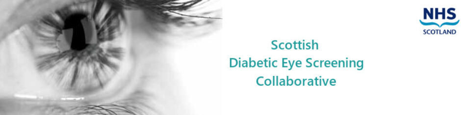
Unlike patients with atrophic age-related macular degeneration, this form can be associated with rapid central visual loss. Typically patients complain that objects are distorted. Sometimes they complain that objects also appear smaller than usual. Both symptoms are a result of the photoreceptors being pushed out of their normal alignment. The cardinal sign is the presence of retinal elevation as a result of fluid leaking from the new vessel and the retina. This is often accompanied by haemorrhage and exudate formation.
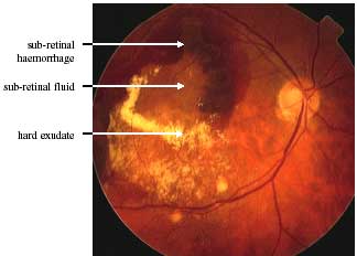
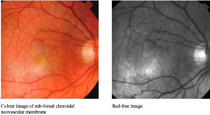
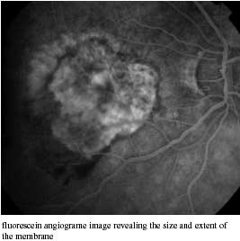
Unfortunately, these new blood vessels often grow under the fovea, very centre of sight. This makes them very difficult to treat. Occasionally they can be treated with ablative laser or with photodynamic therapy. To be effective, treatment must be started before too much vision has been lost.
Untreated, the end result is often a scar. This is termed a disciform scar because of its shape.

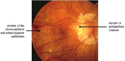
Pathological Myopia
Pathological myopia is characterised by a refractive error of at least -6 diopters and an axial (cornea to retina) length of >26mm. Progressive scleral thinning leads to “expansion” (a form of ectasia termed staphyloma) of the globe. This in turn, results in chorioretinal stretching which manifests as:
- Peripapillary atrophy (optic disc crescent)
- Lacquer cracks (rupturing of Bruch’s membrane)
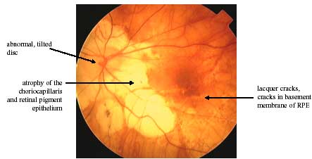
Chorioretinal atrophy associated with overlying vitreous abnormalities. Another common abnormality is tilting of the optic disc.
Patients with pathological myopia are at increased risk of:
- choroidal neovascular membrane formation
- retinal detachment