
D WHAT IS THE NORMAL APPEARANCE OF THE EYE?
1 External Appearance
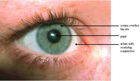
The human eye is a sphere, 2.5cm in diameter with three main layers. The outer supporting coat consists of the transparent cornea, which is continuous with the opaque sclera. The middle layer is the vascular uveal tract and consists of the choroid, ciliary body and the iris, which has a central opening, the pupil.
The inner layer consists of the retina, which can be divided into two- the neurosensory layer containing the photoreceptors which “capture the image” and a layer of pigmented (“brown”) highly metabolically active cells (retinal pigment epithelium) that nourish the photoreceptors.
The contents of the eye consist of the lens and a “jelly-like” substance- the vitreous gel. The lens, along with the cornea, focuses the light onto the centre of the retina. It is suspended by “zonules” that are attached to the ciliary body. This arrangement allows the lens to be “stretched” or “shortened” to enable focusing on objects close to the eye. The centre of the lens is called the nucleus. The nucleus is surrounded by cortex and the whole lens is contained within a close fitting “bag” called the capsule.
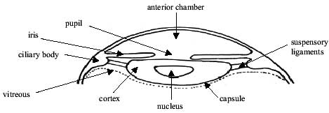
The anterior chamber is the space between the cornea and the lens. The posterior chamber is the space between the lens and the front of the vitreous.
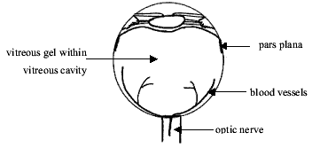
Most of the eye is filled with a “jelly-like” substance, the vitreous gel, which lies behind the lens. It is sometimes likened to a pear. Its base is firmly attached to the back of the eye just beyond the ciliary body. Its stalk is firmly attached to the optic nerve. Elsewhere it is less firmly attached to blood vessels on the surface of the retina.